To accurately assess and characterize the external nose, we have to obtain a solid understanding of the underlying anatomy. In fact, multiple structures influence the surface topography of the nose including the skin (varies in thickness along the nose) and the underlying structural framework of bone and cartilage (osseocartilaginous skeleton).
Since this is a large topic, I will focus on the skin and subcutaneous tissues here with a future post on the osseocartilaginous skeleton.
Skin
The thickness of the skin is one of the most important factors to assess before rhinoplasty. Skin thickness varies from person to person and even varies along the nose itself. Skin tends to be thinner and mobile in the upper half of the nose and thicker and more adherent in the lower half. It has been shown to be thinnest at the rhinion region which represents the point at which the nasal bones intersect with the upper lateral cartilages. Skin also tends to be thinner along the margins of the ala and in the columella.
Your individual skin thickness is so important that it often places limits on what can be achieved aesthetically with rhinoplasty. Thick skin may not allow you to get the definition you desire while minor irregularities or grafts will readily show through in thin skin.
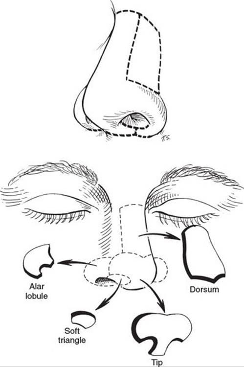
The skin is divided into distinct nasal subunits. These divisions, which are based on the way light and shadow interacts with the nose are extremely relevant in cases of nasal reconstruction. Scars placed at the boundaries of these subunits tend to heal in a more aesthetically pleasing manner. For this reason, it is commonly advocated to remove and reconstruct a whole subunit if there is more than 50% missing either due to trauma or surgical removal.
 Subcutaneous Layer
This is the tissue that lies between the skin and underlying ossecartilagious framework we mentioned earlier. It is made up of four layers –
- Superficial fatty layer (panniculus)
- Connected to the dermis or deep skin
- Fibromuscular layer
- Houses the muscles of the nose
- A part of the SMAS layer of the face which connects the platysma in the neck with the frontalis and galea in the upper face and temporoparietal fascia in the temple.
- Must respect this layer during rhinoplasty to avoid scarring of deeper tissues to the superficial fatty layer that connects directly to skin
- Deep fatty layer
- The major superficial blood vessels and nerves run within this deep fatty layer
- Periostuem and Perichondrium
- This refers to the layers that line the nasal bones and nasal cartilages respectively.
The correct plane for surgical dissection is deep to the deep fatty layer but just superficial to the periosteal or perichondrial layers. An areolar plane exists at this point which parallels that between the galea and periosteum of the scalp.
Nasal Muscles
The muscles of the nose are found in the fibromuscular layer. These have been divided by Drs. Daniel, Griesman, and others into 4 groups based on their function.
- Elevators (shorten the nose and dilate the nostrils)
- Procerus, levator labii superioris alequae nasi, anomalous nasi
- Depressors (lengthen the nose and dilate the nostrils)
- Alar nasalis, depressor septi
- Minor Dilator (dilates the nostrils)
- Dilator naris anterior
- Compessors (lengthen the nose and narrow the nostrils
- Transverse nasalis, compressor narium minor
In certain cases, we can intervene either surgically or with neurotoxins (botox, dysport) to target specific muscles in order to achieve both functional and aesthetic goals.